Self Muscle Massage pt 9- the foot
This is part nine in our self muscle massage series. In the introduction to this series we introduced and demonstrated the three techniques that will be used in this post. If you would like to review them, click here. If you would like to see any other parts of the series, click here.
In the next part of our series, we’re going to be talking about the muscles of the foot. This area is home to four unique layers of muscles that work together to provide movement and shock absorption as they support the joints and arches of the foot during weight bearing and walking. During normal gait, the foot lands on the outside of the heel and must then absorb and transmit that force to the inside of the foot to allow for push off from the big toe. If there is any disruption in this process (due to tight muscles for example), it is very common for the small structures of the foot to break down under the load of the body. Some examples of injuries that can occur include plantar fasciitis, heel spurs, sesamoiditis, neuromas and bunions.
Potential causes of injury:
#1 Abnormal rotation of the foot. If the foot is twisted too much or too little during the weight bearing process (i.e. overpronation or underpronation/supination), the smaller structures of the foot can be injured. This can be caused by lax joints in the midfoot and arches or due to soft tissue restrictions in the long tendons of the lower leg (peroneals/post tib).
#2 Bunion/loss of 1rst MTP extension. In the event that motion becomes restricted in the this area, the foot will become unable to fully load the big toe in preparation for push off. Over time this will lead to compensation and rotation of the lower leg and ankle to allow the foot to fully flatten to the ground during full weight bearing. As the rotation occurs, the gastroc and soleus become less efficient and the smaller muscles of the lower leg must assist with forward propulsion.
#3 Ankle restrictions. These can be the result of sprain/strain injuries where the ligaments, bones, and joints have been affected or from the larger muscles of the lower leg (gastroc, soleus, achilles). If the ankle is restricted, particularly in dorsiflexion (bringing the toes/ankle up towards your nose), the larger muscles will be unable to fully help while walking. This will shift increased work to the foot and big toe to provide push off.
Anatomy
Landmarks
Basic bony landmarks for the foot are easy. You have the inner and outer ankle bone (medial and lateral malleolus) and the heel bone (the calcaneous). Instead of focusing on the million little bones, ligaments and joints in between, we’re going to focus on the structures important to the muscles and key areas we’ll be working on.
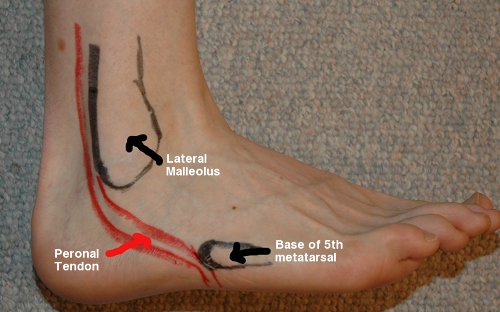
#1 Peroneal Tendon. As you may remember from the last blog post on the outer ankle and shin, the long peroneal tendon runs down the outside of the lower leg, runs behind the ankle bone and to the base of your little toe (5th metatarsal). From there it wraps under the outside of the foot into the arch. To find where that tendon moves beneath the arch start at your little toe. Trace the bone backwards towards your ankle bone. As you get to the middle of the foot you should feel a small bump and the bone will drop off (become less superficial). This is the base of the 5th met. and directly behind it is where the peroneal tendon passes through. If you can find this bump, you’ll be to work on the tendon as it moves towards the arch.
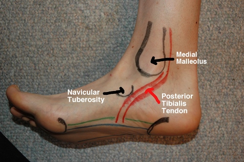
#2 Post Tib Tendon. On the inside of the ankle, you will find the second long tendon that we’ll talking about- the posterior tib. This tendon runs from deep in the calf, down behind the inner ankle bone (medial malleoulus) and behind the navicular bone before it also wraps underneath the arch of the foot. To find the navicular bone start on the inside of the big toe and trace your finger backwards along the bone (NOT the soft tissue of the arch; it’s above the arch). As you get towards the midfoot area you will find a big bump (it may be visible depending on your feet). You can’t miss it. This is the navicular tuberosity and the posterior tibialis tendon passes behind it and below it to wrap around the arch. If you can find this bump, you’ll be on the right area to work on the tendon.
Muscles
The foot is actually home to four different layers of muscles. On top of those is your planar fascia (sometimes called the plantar aponeurosis).
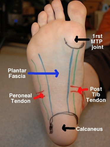
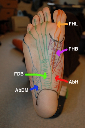
#1 The Plantar Fascia. The PF starts on the heel bone (calcaneous) and then moves up to the ball of the foot and toes (also known as the heads of the metatarsals, one for each toe). It is a thick connective tissue that supports the arch of the foot to provide support during weight bearing.
#2 Post Tib + Peroneal Tendons. On either side of the plantar fascia, you will see the two long tendons of the post tib and long peroneal where they wrap around into the arch. The posterior tib is responsible for pulling the foot in towards midline while the peroneal tendon pulls the foot out away from the body. These are important in determining how the foot functions. If one muscle develops a contracture (chronic shortening of the muscle fibers) it will maintain the foot in a tilted position. This will disrupt how the foot accepts weight and pushes off.
#3 The deeper muscles. Directly underneath the plantar fascia you will find the smaller muscles of the foot. An easy way to visualize these muscles is to start on the heel. There are three main muscles that branch off of the calcaneus. The outermost is your Abductor Digiti Minimi (AbDM). This small muscle controls the little toe and provides support to the outside of the foot during heel strike. Directly next to this muscle is the Flexor Digitorum Brevis (FDB). This small muscle is what pulls the toes down into flexion (curls them). There are four tendons, one each to the smaller toes. Directly next to the FDB is the Abductor Hallicus (AbH). This muscle is the innermost along the arch and is responsible for pulling the big toe out away from the other toes. It wraps around from the inner heel to the outside of the base of the big toe. Once you have found these three muscles, you can move up the foot towards the big toe. In between the tendons of the FDB and the AbH lies the next muscle on our list- the Flexor Hallicus Brevis (FHB). This muscle is responsible for pulling the base of the big toe down into flexion and is unique from the others in that it is home to the two sesamoid bones (the little circles in the purple muscle). The sesamoids are like mini-knee caps for the big toe. They help generate power for propulsion. This muscle runs from deep in the foot to the base of the first metatarsal. It is important to note that this muscle does not go above the 1rst MTP. However, the tendon of the Flexor Hallicus Longus (FHL) does. This muscle is deep in the calf of the lower leg and runs down along the inner ankle before running the length of the foot to the tip of the big toe. This tendon runs between the two sesamoids and is responsible for flexing the full toe.
Soft Tissue Release
What you’ll need: small foam roller or frozen water bottle and a tennis ball.
The techniques:
1) Lengthening/elongation with the foam roller or stick.
2) Cross friction with tennis ball.
3) Sustained pressure or trigger point release with tennis ball.
Key Areas to work on:
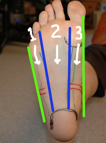
#1 Plantar fascia/FDB + AbDM + AbH. When using the foam roller or frozen water bottle on the bottom of the foot, break the area down into three vertical sections- 1) one from the bottom of the little toe to the heel, 2) one from the bottom of the big toe to the heel, and 3) everything in between to the heel. As you use the roller, be sure to rotate foot to get all three sections. Start with the middle portion in the seated position and progress to standing to increase the pressure. Then roll your foot to the inside to the get that strip. Lastly, roll the foot to the outside to get that strip. This will allow you to get the three main muscle coming off of the heel and lengthen them all the way to where they insert at the ball of the foot under the toes.
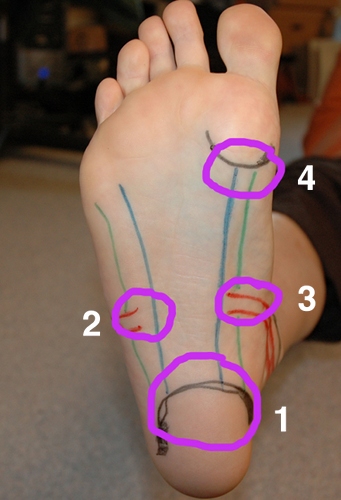
#2 The Tendons. Once you have warmed up the foot with the foam roller you are ready to move onto the deeper techniques of cross friction and trigger point. Remember: the key is to work perpendicular to the tendons when using the cross friction technique. Sink in deep first and then start the movement. It should be very small (approx 1 inch). Key areas that you will want to hit will be along the heel bone (calcaneous). You will want to work all the way around the heel to get the three muscles that originate there. Moving up the foot you will then want to work on the peroneal tendon (#2) and the posterior tib tendon (#3). These come in horizontal so be sure to work in an up and down direction. Lastly, you will want to work just below the big toe. This will get the FHB muscle, as well as, the tendon of the FHL. If you have pain tenderness into the big toe, you can use your fingers to perform cross friction on the tendon. Once you have performed the cross friction technique, you can move onto the sustained pressure/trigger point release. The above 4 areas are common places to hit as well as directly in the middle of the foot for the plantar fascia.
Video
Here is a video demonstration of the soft tissue release techniques of the foot.
Foot from Leigh Boyle on Vimeo.
References:
1) Hammer, Warren. (2007). Functional Soft-Tissue Examination and Treatment by Manual Methods, 3rd edition. Jones and Bartlett Publishers, Inc, Sudbury, MA.
2) Moore, Keith and Dalley, Arthur. (1999). Clinically Oriented Anatomy, 4th edition. Lippincott Williams and Wilkins, Baltimore, MD.



You ROCK!
your video was the best I found,
in terms of some answers to my foot pain,
the information was very simple to understand yet
remained professional in verbage and also very visual in
order for the viewers to participate in the physical therpy.
Thank you,
Tammie Welch