Self Muscle Massage- pt 2 Hamstrings
This is part two in our self muscle massage series. If you missed the last post on the calf or would like to see it again, click here. Also, in the introduction post we discussed and demonstrated the three muscle release techniques that will be used below. Click here if you would like a review.
The next muscle group we’re going to talk about is the hamstrings. Like the gastroc muscle in the calf, the hamstring group crosses two joints (the hip and knee), making it particularly susceptible to injury. In normal gait, the hamstring slows the leg as it swings through, helps stabilize the knee during weight bearing/stance, and then assists with extending the hip through push off. If any disruptions occur during that progression, the hamstring can be overloaded and injured.
To give you some examples of how this might occur:
1) weak quadriceps. following surgery or a knee injury, knee extension is limited during mid-stance and push off. the result is that the hamstrings must work harder to extend the hip during the propulsion phases to make up for the lack of quad contribution. This can also occur in the event of a hamstring contracture (chronic shortening of the muscle) where the knee is unable to fully extend through its’ full ROM.
2) decreased ankle/foot mobility. like the first scenario, this can occur following an injury (such as an ankle sprain), surgery (such as an achilles repair), or in the event of bony abnormalities (bunion, loss of big toe mobility). All of these would subsequently decrease the degree of toe off needed for efficient propulsion. In turn, this would require the hamstring and hip extensors to pick up the slack during push off. This can also occur in the event of calf muscle contractures.
3) decreased hip mobility. In the event of a tight joint or degenerative changes like osteoarthritis, hip extension may be become limited. This would decrease the amount of push off available and shift the work load from the gluteal muscles to the hamstrings/quadriceps. This can also occur in the event of a hip flexor muscle contracture.
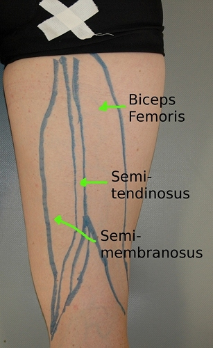
There are three individual muscles: the semimebranosus, the semitendonosis, and the biceps femoris. If you simplify it…all three share the same origin on your ischial tuberosity (sit bone); two come down behind the inside of your knee and the third comes down to the outside. Here are some pics to help give you a visual (this is a right leg).
1) Biceps Femoris (lateral hamstrings). Of the three hamstring muscles, the biceps is the largest and most powerful. It is compromised of a long and short head that both cross the knee joint before inserting onto the tibia.
2) The Semitendinosus and Semimembranosus (medial hamstrings). These two muscles are smaller than the biceps and run down the inside of the back of your thigh. The semitendinosus is a small, thin muscle that has a long tendon, while the semimembranosus is a larger muscle with a shorter tendon. Of these two, the semitendinosus is the most important. This muscle wraps around the inside of the knee to insert into the front of the tibia. When inflamed of chronically tight, it can contribute to pain around the knee cap.
Self Muscle Release Techniques:
What you'll need: a foam roller and tennis/trigger point ball.
The techniques:
1) Elongation/lengthening with the foam roller.
2) Cross friction with the tennis ball.
3) Sustained pressure/trigger point release with the tennis ball.
Key Area’s to work on:
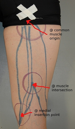
#1) common insertion point at the ischial tuberosity (sit bone).
#2) intersection of all three hamstring muscles. an easy way to find this is to bend your knee and pull your heel into the table. follow the two hamstring tendons up the back of your thigh to where they meet in the middle. move just slightly above that (maybe an inch or two).
#3) the inside tendons above the knee and where they wrap around to the front. due to their ability to contribute to pain around the knee cap, the medial hamstrings should be a point of interest.
Here’s a video to help demonstrate the techniques:
References
1) Hammer, Warren. (2007). Functional Soft-Tissue Examination and Treatment by Manual Methods, 3rd edition. Jones and Bartlett Publishers, Inc, Sudbury, MA.
2) Moore, Keith and Dalley, Arthur. (1999). Clinically Oriented Anatomy, 4th edition. Lippincott Williams and Wilkins, Baltimore, MD.

